Smithsonian
July 19, 2013

Norman Barker was fresh out of the Maryland Institute College of Art when he got an assignment to photograph a kidney. The human kidney, extracted during an autopsy, was riddled with cysts, a sign of polycystic kidney disease.
“The physician told me to make sure that it’s ‘beautiful’ because it was being used for publication in a prestigious medical journal,” writes Barker in his latest book, Hidden Beauty: Exploring the Aesthetics of Medical Science. “I can remember thinking to myself; this doctor is crazy, how am I going to make this sickly red specimen look beautiful?”
Thirty years later, the medical photographer and associate professor of pathology and art at the Johns Hopkins University’s School of Medicine will tell you that debilitating human diseases can actually be quite photogenic under the microscope, particularly when the professionals studying them use color stains to enhance different shapes and patterns.
“Beauty may be seen as the delicate lacework of cells within the normal human brain, reminiscent of a Jackson Pollock masterpiece, the vibrant colored chromosomes generated by spectral karyotyping that reminded one of our colleagues of the childhood game LITE-BRITE or the multitude of colors and textures formed by fungal organisms in a microbiology lab,” says Christine Iacobuzio-Donahue, a pathologist at the Johns Hopkins Hospital who diagnoses gastrointestinal diseases.
Barker and Iacobuzio-Donahue share in interest in how medical photography can take diseased tissue and render it otherworldly, abstract, vibrant and thought-provoking. Together, they collected nearly 100 images of human diseases and other ailments from more than 60 medical science professionals for Hidden Beauty, a book and accompanying exhibition. In each image, there is an underlying tension. The jarring moment, of course, is when viewers realize that the subject of the lovely image before them is something that can cause so much pain and distress.
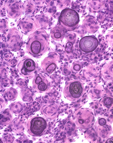
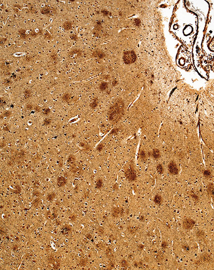
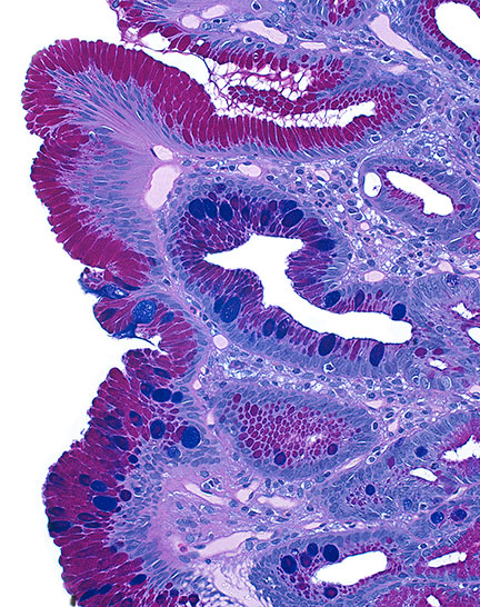
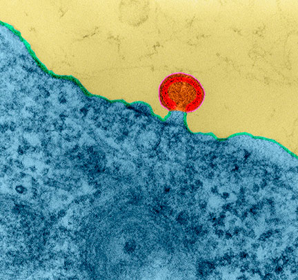
Here is a selection from Hidden Beauty:


